

October
02
/ 2018
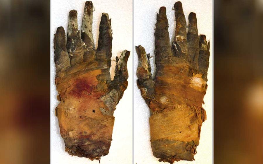
Researchers in Sweden using computed tomography (CT) have successfully imaged the soft tissue of an ancient Egyptian mummy's hand down to a microscopic level, according to a study published in the journal Radiology. Non-destructive imaging of human and animal mummies with X-rays and CT has been a boon to the fields of archaeology and paleopathology, or the study of ancient diseases. Imaging studies have contributed to a better knowledge of life and death in ancient times and have the potential to improve our understanding of modern diseases. Both X-ray and conventional CT take advantage of the fact that materials absorb different amounts of X-rays. This phenomenon, known as absorption contrast, creates different degrees of contrast within an image. "For studying