


CT Scan Conducted on 2,000-Year-Old Mummy Reveals the Cancer that Killed Him
A team of astonished doctors at Crouse Hospital in New York, had the rare opportunity to examine and scan a 2000-year-old mummy. How does it feel to examine a perfectly preserved ancient Egyptian mummy? The hospital’s staff describes it as a once in a lifetime experience.
A Very Unusual “Patient”
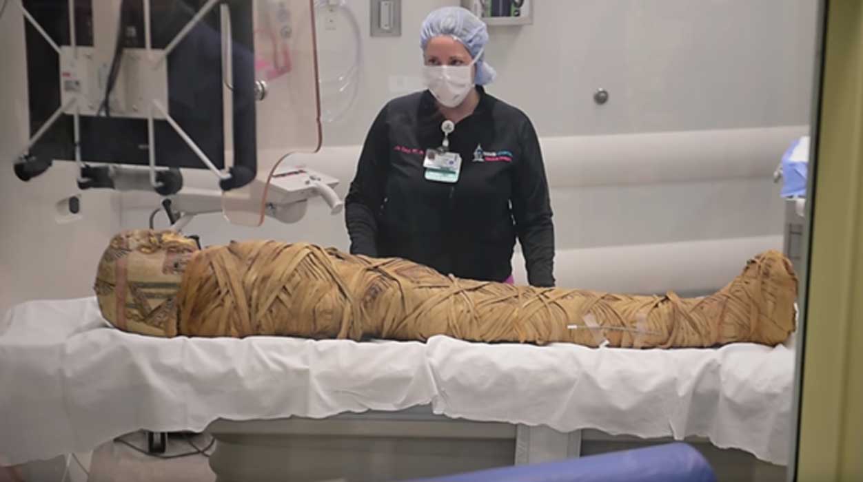
A very unusual patient named Hen, who happened to be a 2,000-year-old Egyptian mummy, was transferred at Crouse Hospital in Syracuse, New York, for a regular CT Scan in order to find out more information about it, on Sunday, December 10. The doctors and nurses working there, couldn’t believe their eyes. "He had a tumor on his Fibula which is one of the two bones of the lower leg," Dr. Mark Levinsohn said according to International Business Times. "Looking at it, it had all the characteristics of a malignant tumor and one that's somewhat rare. So, here we have a rare circumstance and a rare tumor and that evoked our interest a lot," he added.
- CT Scans of Mummy of an Ancient Priest Reveal He Was Stricken with Modern Diseases
- Earthquake in Ecuador Reveals Bizarre Burial of a Mummy in a Jar with a Little Mouse

The mummy is around 2,000 years old and from Egypt. (Youtube Screenshot)
CT Scanning Reveals the Type of Cancer
Doctors were able to identify the exact type of tumor the mummy had (during his lifetime) thanks to the gigantic steps forward medical technology has taken during the past few years. Just eleven years ago, the mummy had been scanned but the less advanced CT scan and biopsy tools at the time didn’t allow doctors to reveal the type of cancer that eventually killed the mummified individual. "Since that time, the last ten years, they've upgraded the equipment. What, at that time was a 16 detector scanner is now a 320 detector scanner and all that additional information is now derived when we scan the body. So, we can tell all kinds of greater detail," Levinsohn told International Business Times.
- New Tech Brings 2,000-Year-Old Mummy of Little Egyptian Girl to Life in Stunning Detail
- First-of-its-Kind Mummy Study Reveals Clues to Girl’s Story

Mummy has now been scanned by a 320 detector CT scanner. (Youtube Screenshot)
The Role Modern Technology in Examination of Mummies
Indeed, as Kingtutone reports, nowadays scientists can examine mummies in a different way than a decade ago, as x-ray analysis, CT scanning, and DNA testing has progressed dramatically. The times of opening a mummy physically have faded into the past and now much of the work is done through advancements in technology. New technological advancements are allowing researchers to peel back the layers digitally, thus, giving us a view of the preservation process without destroying any evidence. The benefits are astounding. Medical information can be determined in a matter of minutes, exposing the mummy fully only takes seconds, and digital records can be saved for further examinations.
- Scans and DNA tests reveal the secrets of a rare African mummy
- Mystery wrapped in linen: Unraveling the story of Hatason, a 3,200-year-old Egyptian mummy

The more detailed CT scan revealed the mummified individual had a tumor on his fibula. (Youtube Screenshot)
Especially CT scanning helps scientists to study the bodies without disturbing its contents and allows scientists to see things exactly as they were 2000 years ago. By taking pictures of the mummies in layers, soft tissue such as the organs can now be seen untouched and unaltered. Several digital images are combined together recreating the body in 3D. This allows clear viewing of what’s beneath the wrappings and shows everything as it was thousands of years ago.
No matter how advanced modern technology may be nowadays, bones and teeth still remain an important factor of deciphering the life of a mummy. Through bones and teeth, an individual’s age and cause of death can be predicted. As Kingtutone reports, younger mummies are determined more easily due to the growth in the bones and layers in the epiphyses, which are the joints. The layers help to identify the body’s age up until 25 when all the layers combine together. Bones also reveal the quality of life, certain diseases and injuries throughout the mummy’s life.
Hen was initially brought to the Cazenovia Library and Museum in New York by Robert Hubbard in 1894 from Egypt as Spectrum News reported, and will soon be returned to the controlled atmosphere environment there where he resides.
Top image: The mummy ‘Hen’ has been sent for a hospital CT scan for the second time. (Youtube Screenshot)
